Earth-Sciences Electron Microscopy and in-situ X-ray Microanalysis
(formerly SEM – microprobe laboratory)
School of Earth Sciences, The University of Melbourne, Parkville Level 1, room 131
Mr Graham Hutchinson
Laboratory manager and booking contact
Dr Andrea Giuliani
Laboratory supervisor
The laboratory houses
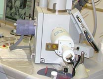
Philips FEI XL30 environmental scanning electron microscope (ESEM) equipped with an OXFORD INCA energy-dispersive X-ray spectrometer (EDS) and a Gatan cathodoluminescence (CL) detector*.
The main aims of our SEM are to provide high-resolution images (up to ~50,000x) of geological samples for micro-textural and micro-morphological studies and to identify mineral phases in thin sections and mounts through semi-quantitative major-element chemical analyses. The SEM employs an electron beam to excite the surface of the exposed sample. Interaction of the exposed surface with the electron beam produces a variety of signals:
- backscattered electrons (BSE) reveal differences in average atomic number visualized as brightness variations on the microscope display screen – heavier minerals have higher brightness and vice-versa. Flat, polished and carbon-coated thin sections/mounts are preferable;
- secondary electrons (SE) show surface morphology;
- cathodoluminescence (CL) reveals trace element variation in chemistry and internal growth patterns/stresses (only certain minerals exhibit cathodoluminescence);
- X-rays are detected by the energy dispersive spectrometer (EDS) for semi-quantitative major-element chemical analyses. Flat, polished and carbon-coated thin sections/mounts are preferable. Our SEM produces single-spot chemical analyses as well as 2D maps of the major elements.
We prefer to work on carbon- or gold-coated samples to minimize instrument exposure to micro-particles residing on sample surfaces and introduction of aqueous vapour in the system, both of which can damage the electron column; however, in special circumstances, we may be able to accommodate short sessions on uncoated samples (please direct enquiries to Graham Hutchinson in advance);
Cameca SX50 electron microprobe (EMP) equipped with four vertical wavelength dispersive spectrometers (WDS)**.
The EMP provides quantitative major-element chemical analyses of mineral phases and other materials in thin sections and mounts. Analyses of single spots and spots along traverses are possible. The thin sections and mounts must be polished and coated with carbon (or gold) for EMP analysis. The phase to be analysed is excited by a finely focused (>1-2 μm) electron beam and the X-rays produced are detected by the wavelength dispersive spectrometers (WDS). A defocused beam can be utilised to minimise volatilisation of alkali and volatile elements (e.g., K, Cl, F, etc.) for minerals like micas. Detection limits vary from element to element but are generally at or below ~300 ppm (~0.03 wt.%) when standard settings are employed. The points for analysis are pre-selected by the customer using an optical microscope, which is available in the facility and equipped with a Microbeam Digimax digitiser system. Two small (~3 mm) grids are located on each thin section/mount as reference points for the points subsequently selected by the customer. The operator then performs the analyses and delivers the results to the customer. Alternative arrangements can be discussed for special circumstances (enquiries to Graham Hutchinson in advance);
ZEISS petrographic microscope equipped with 5 objectives (5, 10, 20, 50, 100x).
A computerised stage with a remote joystick controller and an OLYMPUS camera for low-resolution photos of the phases selected for EMP analysis. A Microbeam Digimax digitiser system is attached to the microscope to select and store points for EMP analyses.
The Digimax files can be easily converted to CSV files and the points can be utilised by other instrumentation
- e.g., for example for laser-ablation ICP-MS analysis;
- a ZEISS binocular stereoscopic microscope;
- a JEOL JEE 4C carbon coater with resistive carbon thickness monitoring;
- an EMITECH K950X Turbo Evaporator equipped with a thickness detector for gold coating.
FEI Quanta 600 MLA environmental scanning electron microscope (ESEM).
This instrument will replace the Philips FEI XL30 ESEM, which will be employed to work on non-coated samples. More details soon. ** In 2015 a new electron microprobe will be commissioned and it is expected to be fully operational by the end of 2016.
Please contact Graham Hutchinson for further enquiries and instrument booking.
Detailed instrument configuration
Philips FEI XL30 ESEM
- Installed 2002
- Tungsten source
- Vacuum system utilises an air-cooled turbomolecular pump (TMP) and two rotary vane pumps
- 2 vacuum modes: high vacuum, ESEM low vacuum (up to 1 torr)
- FEI detectors: Everhardt-Thornley SE, clip-on 2-segment solid state BSE, CCD Camera for chamber observation
- Gatan PanaCL panchromatic CL detector with RGB filters
- Oxford INCA EDS system with UTW Si(Li) detector, MnKα 132 eV resolution
- 5-axis motorised stage, XYZ range 50x50x65mm
Cameca SX50 EMP
- Installed May 1989, serial number 239
- Tungsten electron source; 50kV maximum accelerating potential
- 4 optically encoded WDS spectrometers with LiF, PET, TAP, and PC0 analysing crystals covering elements from boron to uranium
- PC computer control by SAMX Xmas software package
- Cameca SE detector
- Optical microscope (reflected/transmitted light, polariser)
- 3-axis motorised stage, XYZ range 55x80x2mm, 1um repeatability, optical encoders
- Ion getter pump for long filament life, oil diffusion and rotary vane pumps